
Aedes vexans
Blood-feeding insects often vector pathogens of medical and veterinary concern; as such, the feeding mechanism of these insects deserves further study. Mosquitoes are especially of interest due to the public health importance of diseases they vector. Microscopic examination of the morphology and location of sensory structures (sensilla) within the mouthparts, specifically within the food canal, cibarium (pumping apparatus), and stomodaeum (true mouth), may reveal functionality of these structures.
Sensilla of the cibarium, beginning
at the base of the labrum and extending proximally through the stomodaeum, were
examined in both female and male Aedes
vexans (Meigen) mosquitoes. Two types of sensilla were observed: setiform
and basiconic. Both forms of sensilla were aggregated in the distal region of
the cibarium surrounding a hard palate, with the basicones medially positioned
and the setiforms laterally. Additionally, two pairs of basicones were
consistently observed in the proximal end (stomodaeum) of the cibarium in every
individual examined.
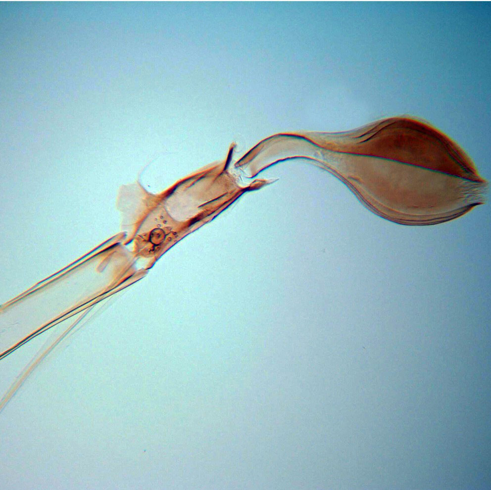
Feeding Structures
When a [female] mosquito bites, blood flows up the labrum into the cibarium, which has an aggregation of sensilla – both basiconic and setiform – in the anterior region. The cibarium is the first of two pumps that allow the mosquito to ingest a blood meal. There are two pairs of basiconic sensilla between the cibarium and the pharyngeal pump, the second (and larger) pumping apparatus. *Male mosquitoes do not take blood meals, but rather feed on plant nectar.
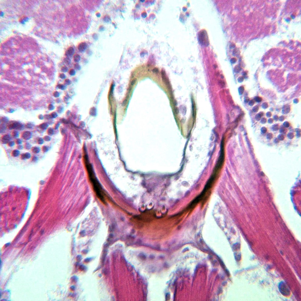
Tissue Sectioning
In order to locate the different types of sensilla within the cibarium, 6-µm tissue sections were obtained and stained with hematoxylin and eosin. With this method, sclerotized structures, sensilla, striated muscle, and ganglia were able to be differentiated. Tissue sectioning was performed by the Histology Department of Cabell Huntington Hospital Laboratory.
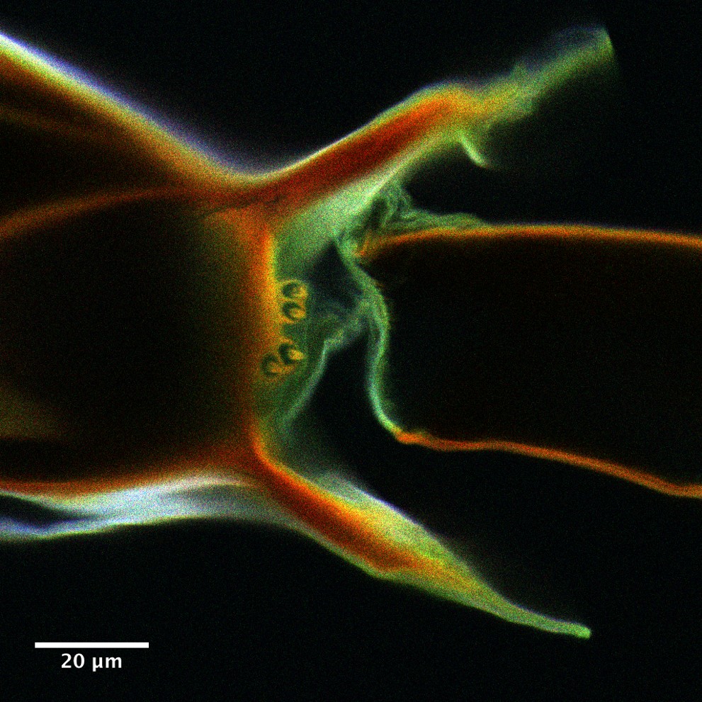
Autofluorescence
Many insects, including mosquitoes, exhibit autofluorescence due to the chitin-content of insect cuticle. As a result, confocal scanning laser microscopy (CSLM) can provide real x, y, and z-data without the use of dyes or fluorescent markers. Three-dimensional projections of sensilla within the cibarium were produced using this method.